PROJECT OVERVIEW
CytoTrack technology is a new kind of scanner that is specifically developed for the challenges of scanning and analyzing rare cells, such as Circulating Tumor Cells (CTC).
The basic idea behind CytoTrack is closely related to conventional CD/DVD technology. The sample is spread on a special glass disc (CytoDisc™). The area of the disc is much larger than a conventional microscope slide and can accomodate up to 100 million cells in a monolayer. This makes it possible to scan all nucleated cells in a blood sample, and hereby eliminate the need for a pre-selection step.
INFORMATION
- Category Partners
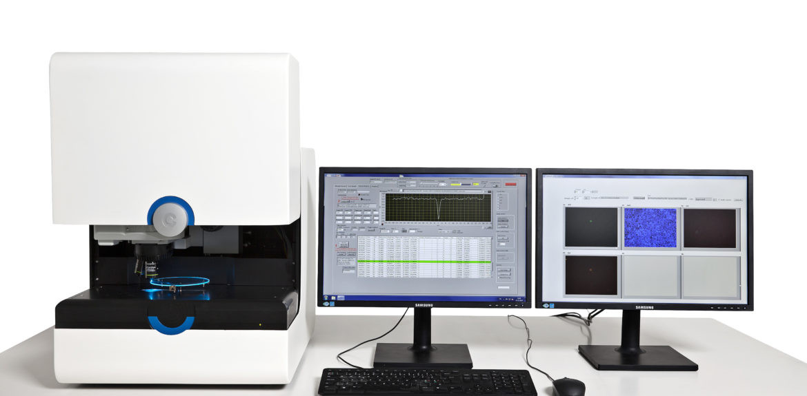
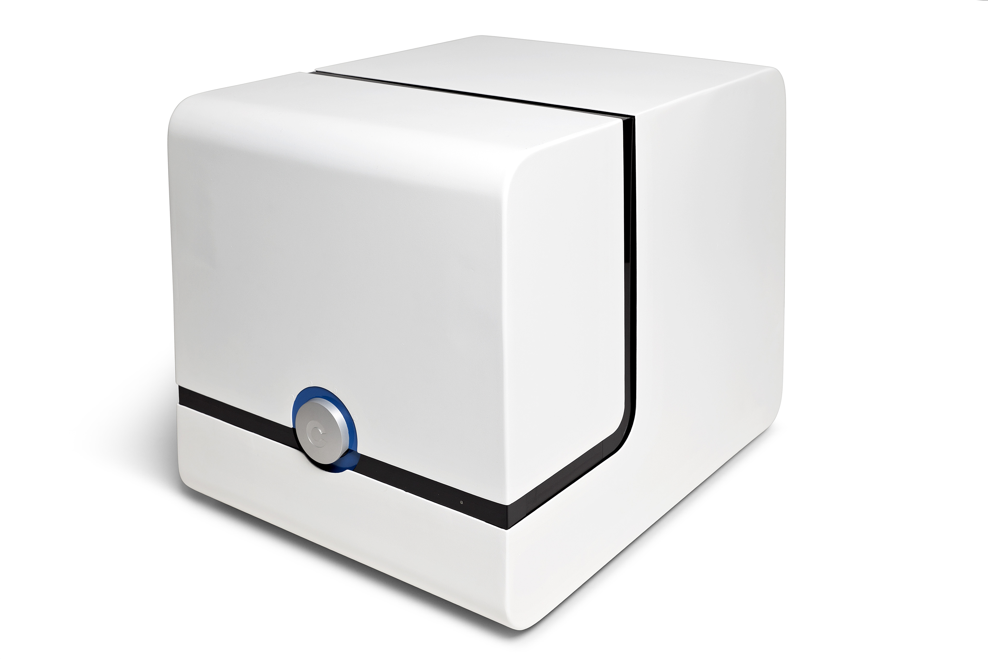
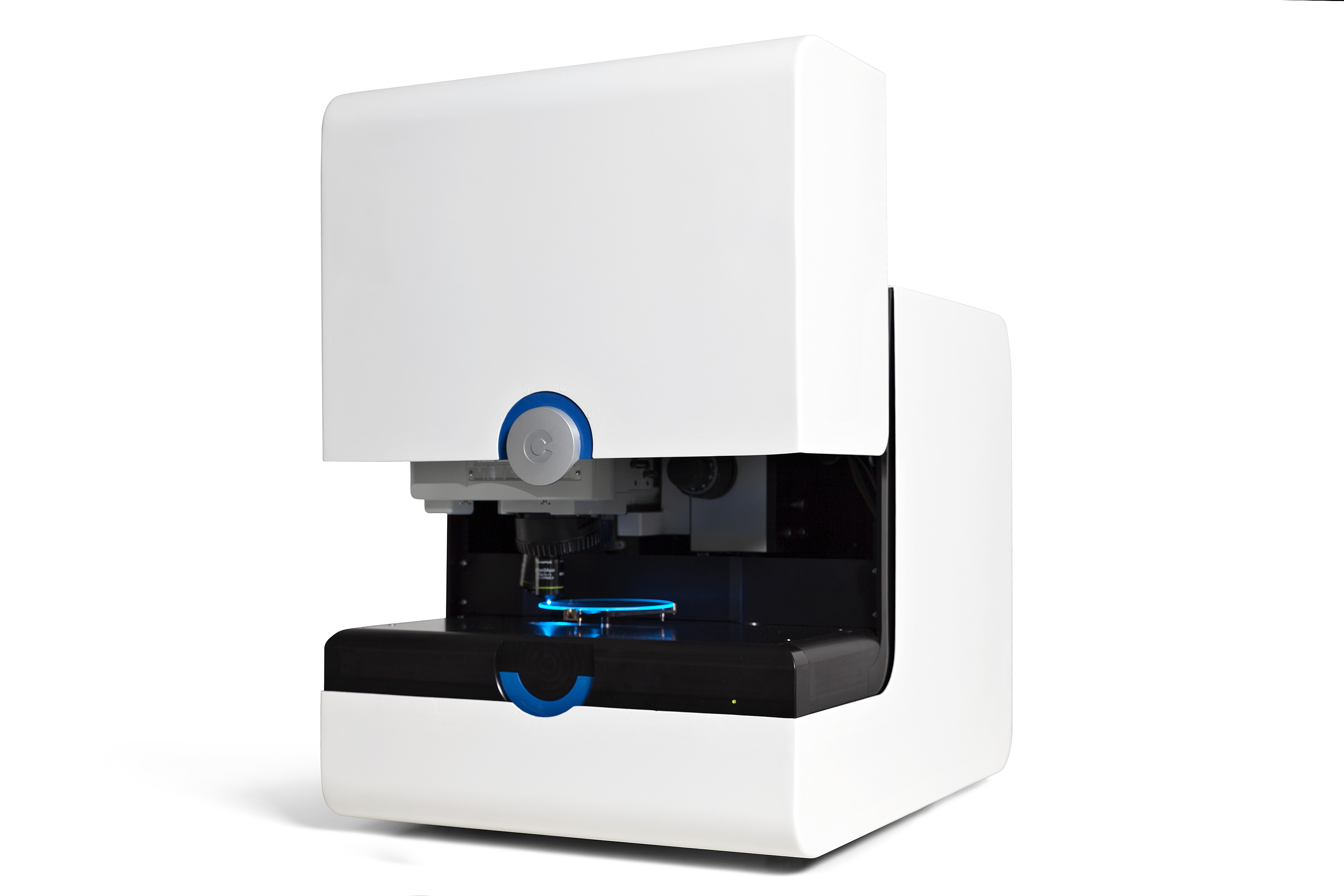
CytoTrack CT11™ scanner
The new CytoTrack CT11™ provides state-of-the art rare cell scanning and isolation capabilities.
This instrument can detect rare cells (such as CTCs) efficiently and at great speed. The optical sensitivity is extremely high.
Individual target cells can be characterized ‘on disc’ or ‘off disc’.
This highly flexible technology is ideal for exploring new possibilities within CTC approaches and techniques.
CytoTrack CT11™ allows users to experiment with different protocols, staining techniques, antibodies and set-ups, thus making it possible to study a variety of cancer types.
CytoPicker™
The CT11™ instrument features the new cell picking functionality. After enumeration of CTCs, selected cells can be lifted from the glass surface and entered in to Eppendorf tubes. Target cells can be isolated completely with no white blood cell contamination, and target cells are kept intact.
Easy to use
The CT11™ has an easy-to-use graphical user interface. The user will be able to perform scans after just a few hours of training.
Each target cell (hot spot) on the disc can be revisited very quickly in less than one second (going from hotspot to hotspot). This makes it very easy to step through a list of targets in order to examine each target cell in detail.
High quality images
The camera provides high resolution (HD images) of the target cells.
The camera has an automatic focusing feature and images are displayed automatically in an overview image gallery.
Some of Key features
01
All nucleated cells in the blood sample are scanned. Due to the extremely high scanning capacity of the CytoTrack scanner, the need for pre-selection is eliminated. No need for a pre-selection step based on EpCAM (magnectic separation) or filtration(based on cell size).
02
After enumeration, selected target cells can be molecular characterized. Either ‘on-disc’ by IFA (fluorescent antibodies) and FISH (fluorescence in situ hybridization). Or ‘off-disc’ by isolating the target cells and performing single cell PCR or sequencing.
03
Individual intact cells can be picked up with the CytoPicker™. After enumeration scanning, the operator can select specific cells to pick up from the disc, and place them into PCR tubes.
04
The CytoTrack detection principle has an extremely high scanning capacity. The scanning principle is comparable with flowcytometry. Cells are scanned optically for the fluorescent signal. After the first scan, each ‘hotspot’ on the CytoDisc™ is revisited and further analysed by imaging. The instrument automatically images each ‘hotspot’ and pictures are shown in an image gallery. This makes it easy for the operator to select CTCs from debris.
05
Due to the ultra high scanning capacity, a pre-selection of cells by filtration is not necessary. The scanning principle is solely based on fluorescent signals, the bias of size is therefore eliminated. Many CTCs have the same size as white blood cells.
06
The instrument is build of the highest quality optical components and provides HD (high definition) images of each individual cell. This makes it easy for the operator to determine the morphology of the cell and staining characteristics.